Findings:
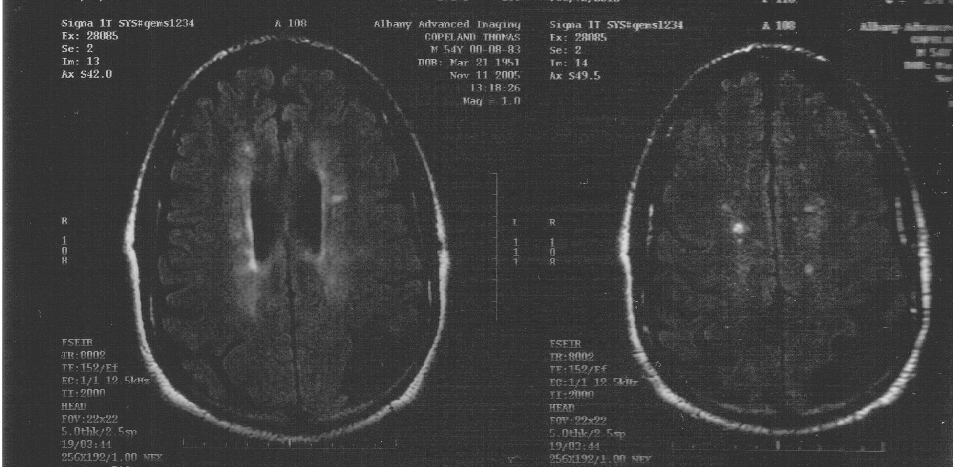
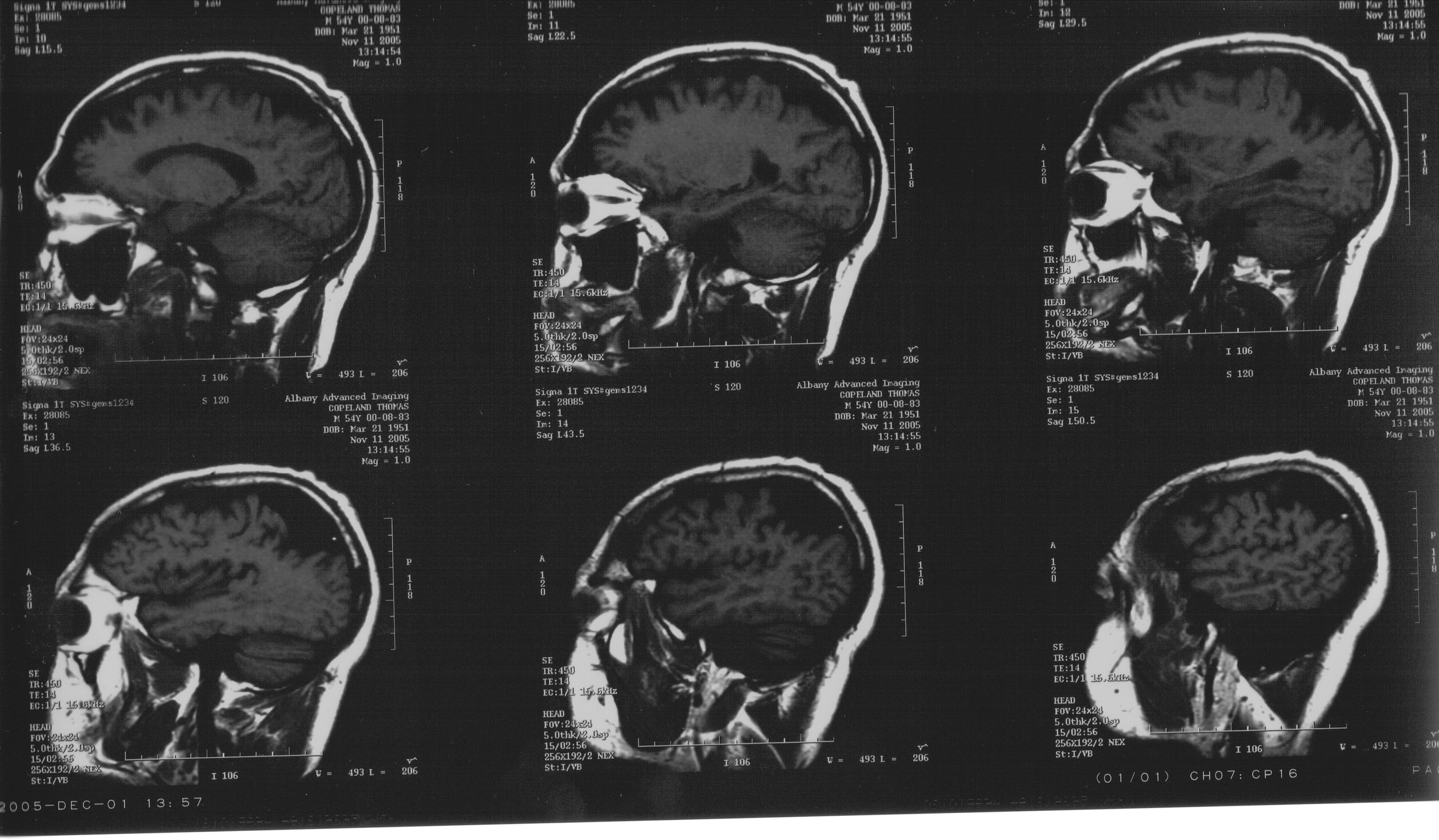
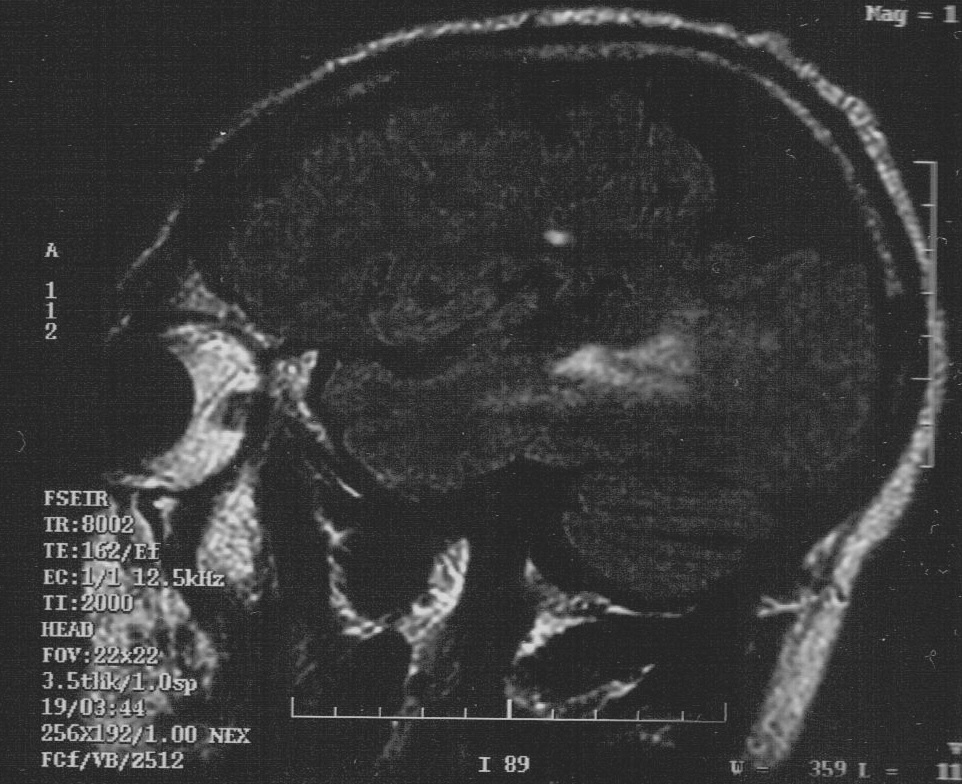
- The Ventricles are at the upper limits of normal in size or slightly enlarged. There is focal Atrophy in the Posterior Parietal Lobes bilaterally. These findings are unchanged compared to previous study. (View: My MRI image)
- Multiple oval areas of increased signal are noted on FLAIR and T2 weighted images in the White Matter of both Cerebral Hemispheres. (View: My MRI image)
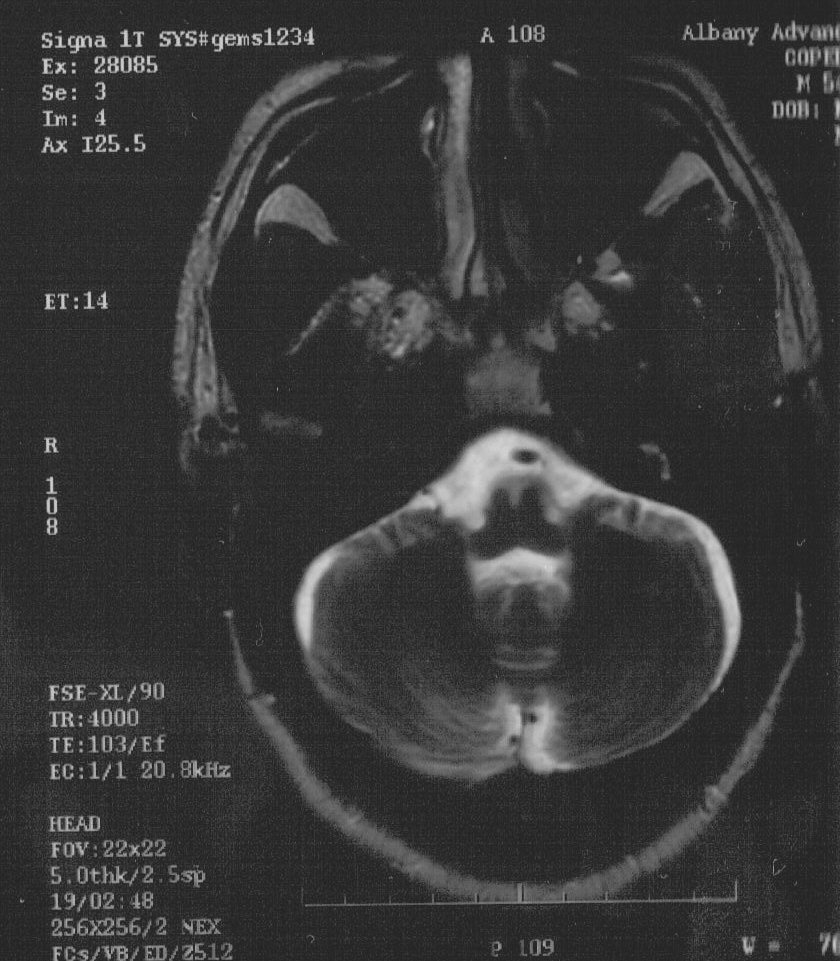
- Since the previous MRI scan, a 6-7mm area of HyperIntensity has developed in the Ventral Pons to the right of midline, seen on FLAIR and T2 weighted images, with restricted Diffusion.
- There is no evidence of abnormal contrast enhancement associated with the Right Ventral Pontine abnormality.
- New punctate T2 signal abnormalities are noted in the left Cerebellar Hemisphere (View: My MRI image)
- There is linear enhancement in the Left Parietal Lobe following Gadoteridol contrast administration, and there may be minimal rounded enhancement adjacent to the body of the Left Lateral Ventricle as well. (View: My MRI image)
- These areas of enhancement are not tightly correlated with areas of T2 abnormality.
- There is no evidence of mass effect or ExtraCerebral Fluid collection.
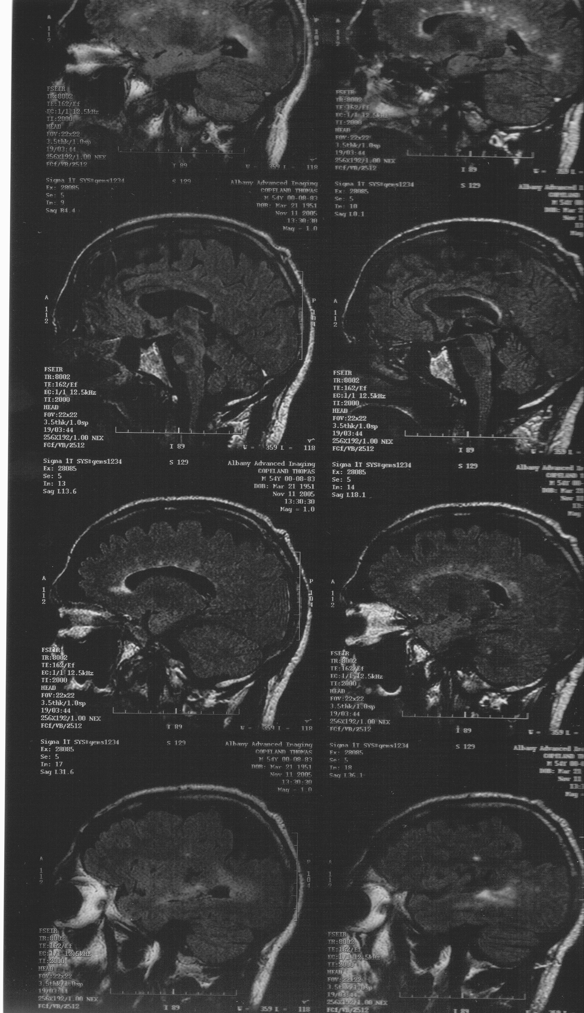
- There is Atrophy of the Corpus Callosum. (View: My MRI image)
Impression:
- Mild Atrophy and White Matter signal changes in both Cerebral Hemispheres, compatible with the clinical diagnosis of Multiple Sclerosis.
- A 6-7mm area of T2 HyperIntensity with restricted Diffusion has developed in the ventral aspect of the Pons to the right of midline. This may well represent a recent, active plaque.
- Linear area of signal enhancement in the left Parietal White Matter. (View: My MRI image)
- While this is not tightly correlated with signal abnormality in this location on FLAIR and T2 weighted sequences, this could represent an additional area of acute DeMyelination.
- Stable focal Atrophy involving both posterior Parietal Lobes, left more than right. (View: My MRI image)