Medical Terminology

#13
1978

by: Dr. James Brevan

p62

The MidBrain & BrainStem
This Vital Control area of the Brain maintains all Essential regulatory mechanisms of the body [The Automatic Nervous System (ANS)].

The Pons
Connects the BrainStem with the balancing control of the Cerebellum.

The Reticular Formation
Links the vital centers with each other and monitor the information reaching the BrainStem from the Sensory Tracts of the Spinal Cord.


#14
1977

*Gray's Anatomy*

Corpus Callosum
A thick band of more than 200 million Transverse Nerve Fibers, which is the only connection between the Hemispheres of the Cerebrum. It lies at the bottom of the Cerebral Longitudinal Fissure.

Cerebral Interior

p656

The Rostrum
The reflected anterior portion, called the Beak or Rostrum. At its termination the Corpus Callosum passes backwards from its under surface, across the Posterior Margin of the Anterior Perforated Space to the Hippocampus Bands - the Peduncles of the Corpus Callosum.

p660

The underside of the Corpus Callosum forms the Roof of the Lateral Ventricles.

The part of the Corpus Callosum which curves forward on each side from the Genu into the Frontal Lobe and covers the front part of the Anterior Cornu of the Lateral Ventricle is called the Forceps Anterior or Minor.

The part which curves backwards from each side of the Splenium into the Occipital Lobe called the Forceps Posterior or Major.
(View: My 6th MRI image)


#15
1986

*Merriam Webster's Medical Desk Dictionary*

p563

Pons: A broad mass of chiefly Transverse Nerve Fibers conspicuous on the Ventral Surface of the Brain. Located at the Anterior End of the Medulla Oblongata.

Pontine Nucleus: Any of various large groups of Nerve Cells in the Basal part of the Pons, that receive Fibers from the Cerebral Cortex and send Fibers to the Cerebellum by way of the Middle Cerebellar Peduncles.

Peduncle: A band of White Matter joining different parts of the Brain.

p592

Punctate: Characterized by dots or points, a structure or area.


Cerebellum

A large Dorsally Projecting part of the Brain that is especially concerned with the Coordination of Muscles and the maintenance of bodily Equilibrium, situated between the BrainStem and the back of the Cerebrum.

Cerebellar Peduncle

Any of three large bands of nerve fibers that join each Hemisphere of the Cerebellum with the parts of the Brain below and in front.

  • The Superior Cerebellar Peduncle: connects the Cerebellum with the MidBrain (Image)

  • The Middle Cerebellar Peduncle: connects the Cerebellum with the Pons (Image)

  • The Inferior Cerebellar Peduncle: connects the Cerebellum with the Medula Oblongata (Image)

CerebelloPontine Angle: A region of the Brain at the junction of the Pons and the Cerebellum, that is a frequent site of Tumor formation.

Cerebral Peduncle: Either of two large bundles of nerve fibers passing from the Pons forward and outward to form the main connection between the Cerebral Hemispheres and the Spinal Cord (CorticoSpinal Tract).


#11
1992

Anatomy & Physiology

2nd Edition

by: Rod R. Seeley, Ph.D.
Trent D. Stephens, Ph.D.
Philip Tate, D.A.


Basal Ganglia

p397

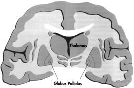
The Basal Ganglia are a group of functionally related Nuclei located bilaterally in the Inferior Cerebrum, DienCephalon and MidBrain. The SubThalamic Nucleus is located in the DienCephalon, and the Substantia Nigra is located in the MidBrain.

The Nuclei in the Cerebrum are collectively called the Corpus Striatum (Stripped Body) and include the Caudate Nucleus (Tail) and Lentiform Nucleus (Lens Shaped). They are the Largest Nuclei of the Brain and occupy a large portion of the Cerebrum.  (View Image)

The Basal Ganglia play an important role in Planning and Coordinating Motor Movements and Posture. Complex neural connections link the Basal Ganglia with the Cerebral Cortex. The major effect of the Basal Ganglia is to Inhibit Unwanted muscular activity and disorders of the Basal Ganglia result in exaggerated, uncontrolled movements.


The Limbic System

p398

Portions of the Cerebrum and DienCephalon are grouped together under the title Limbic System. The term Limbic is not precise and is used differently by various authors. The term Limbus means Border, and the term Limbic refers to the Medial Portion, or Border of the Temporal Lobe.


Limbic System Structure

  • View Limbic 1   Limbic 2

  • Certain Cerebral Cortica areas, including the Cingulate (To Surround) Gyrus, located along the Inner Surface of the Longitudinal Fissure just above the Corpus Callosum, and the Hippocampus

  • Various nuclei such as Anterior Nuclei of the Thalamus and the Habenular Nuclei in the EpiThalamus

  • Parts of the Basal Ganglia

  • The HypoThalamus, especially the Mamillary Bodies

  • The Olfactory Cortex

  • Tracts connecting the various Cortical and Ganglia (the Fornix is one such Tract)

The Limbic System influences Emotions, the Visceral Responses to those emotions, Motivation, Mood, and sensations of Pain and Pleasure. One of the major sources of sensory input into the Limbic System is the Olfactory Nerves. The smell of food stimulates the Hunger Center in the HypoThalamus.

Lesions in the Limbic System can result in Voracious Appetite, increased (often perverse) sexual activity, and docility (including loss of normal fear and anger responses). Since the Hyppocamus is part of the Temporal Lobe, damage to that portion of the Limbic System can also result in a Loss Of Memory.

The Hyppocamus and the adjacent Cortex are very important in the transition of information from Short to Long Term Memory, and the cells undergoing Calcium induced shape changes associated with Long Term Memory are localized in that region of the Brain.   (Also See: Limbic System


Cerebellum

p400

The term Cerebellum means Little Brain. It communicates with other regions of the CNS through Three Large Nerve Tracts: the Superior, Middle, and Inferior Cerebellar Peduncles.

The Cerebellum is organized like the Cerebrum, with Gray Matter both on the inside as Nuclei and on the outside as Cortex. The Cerebellar Cortex has Gyri and Sulci, but the Gyri are much smaller than those of the Cerebrum.

The Cerebellum consists of Three Portions: the FlocculoNodular Lobe. A narrow Central Vermis, and Two large Lateral Hemispheres.

The FlocculoNodular Lobe is the simplest portion of the Cerebellum and is involved in Balance. The Anterior Portion of the Vermis is involved in Gross Motor Coordination, and the Posterior Vermis and Lateral Hemispheres are involved in Fine Motor Coordination - producing smooth, flowing movements.


A major function of the Cerebellum is that of a Comparator. Impulses from the Motor Cortex descend into the Spinal Cord to initiate Voluntary Movements, and at the same time impulses are sent from the Motor Cortex to the Cerebellum, giving the Cerebellar Neurons information representing the Intended Movement.

Simultaneously, Impulses from the Proprioceptive Neurons (providing information about the Position of the body or body parts) that innervate the joints and tendons of the structure being moved, reach the Cerebellar Cortex (Proprioception).

These impulses give the Cerebellar Neurons information from the Periphery about the Actual Movements. The Cerebellum Compares Impulses from the Motor Cortex with those from the Moving Structures (it compares the intended movement with the actual movement).

And if a difference is detected, the Cerebellum sends Impulses to the Motor Cortex and the Spinal Cord to correct the discrepancy. The result is smooth and coordinated movements.
(Also See: Proprioception)

With training a person can develop highly skilled and rapid movements that are accomplished more rapidly than can be accounted for by the Comparator Function of the Cerebellum.

In these cases the Cerebellum can "learn" highly specialized Motor Functions through specific, repeated comparator activities.


Continued In scan-02a.html


Medical Texts
Anatomy | Immune System | Lymphocytes | Meds
MHC | Movement | Cranial Nerves | Physiology


MS Glossary ThJuland's MSers' Glen - Our CyberHome Page Top The Glen's Gallery: Come & Share Our Stories MS Files MS Abstracts Site Index


Abstracts
ANS | Bladder | Cognition | Fatigue | Fluid | Genetics
Interferons | IVIG | Nitric Oxide | Optic Neuritis | Pain
Physiology | Prions | Prognosis | ReMyelinate | Steroids
Stress | Treatments | TNF | Uric Acid | Viruses


© Copyright 1997 - 2011:
Permission is granted to MS Societies and all MSers to utilize information from these pages provided that no financial reward is gained and attribution is given to the author/s.