#2
Diffusion Anisotropy In The Corpus Callosum
Chepuri NB, Yen YF, Burdette JH, Li H, Moody DM, Maldjian JA
AJNR Am J NeuroRadiol 2002 May;23(5):803-8
Wake Forest University School of Medicine, Bowman Gray Campus, Department of Radiology, Medical Center Boulevard, Winston-Salem, NC 27157, USA
PMID# 12006281
Abstract
Background And Purpose
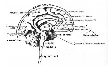
The Corpus Callosum is a heterogeneous White-Matter Tract that connects the Cerebral Hemispheres. The purpose of this investigation was to study its MicroStructural architecture in normal human adult Brains by using Diffusion Tensor imaging (DTI).
Methods
Two hundred consecutive patients referred for Brain MR imaging underwent additional DTI with a high gradient field strength applied in six directions.
Forty-two patients met the following inclusion criteria: 1) normal Brain and 2) age greater than 18 years. Anisotropy maps were generated, and regions of interest were drawn around specified regions within the Corpus Callosum.
Results were stratified by sex and age. In addition, available Histologic specimens of the Corpus Callosum from cadaver Brains were analyzed with conventional and specialized Vascular staining.
Results
Anisotropy values in the various regions of the Corpus Callosum differed significantly. Average values of the Anisotropy index for the Genu, Body, and Splenium of the Corpus Callosum were 0.400, 0.456, and 0.539, respectively.
The differences between these values are statistically significant (P < .01). Increased Anisotropy was present in posterior areas in both sexes and in all three age groups examined.
Conclusion
The results of this investigation show a statistically significant increase in Anisotropy of the Corpus Callosum in its more posterior portions compared with its more anterior portions across sex and age groups.
Although the MicroStructural Etiology for this apparent increase in Anisotropy is unclear, a number of possible mechanisms are presented.
#3
Preferential Occult Injury Of Corpus Callosum In Multiple Sclerosis Measured By Diffusion Tensor Imaging
Ge Y, Law M, Johnson G, Herbert J, Babb JS, Mannon LJ, Grossman RI
J Magn Reson Imaging 2004 Jul;20(1):1-7
New York University School of Medicine, Department of Radiology, New York, New York 10016, USA
PMID# 15221802
Abstract
Purpose
To investigate the feasibility of Diffusion Tensor Imaging (DTI) assessment of microscopic Fiber Tract Injury in the Corpus Callosum (CC) and other Normal-Appearing White Matter (NAWM) in patients with early Multiple Sclerosis (MS).
Materials And Methods
DTI was performed in 12 healthy volunteers and 15 patients who have relatively short disease duration (mean = 2.7 years).
Both Fractional Anisotropy (FA) and Mean Diffusivity (MD) were obtained in different regions of Normal-Appearing CC (NACC) and NAWM in Frontal and Occipital regions.
Results
The data showed significantly lower FA (P < 0.001) and higher MD (P < 0.04) for NACC regions.
But not for Frontal and Occipital NAWM regions, in patients than in those in healthy volunteers after Bonferroni adjustment.
The increase of MD in the entire NACC regions was correlated with the total Cerebral lesion volume (r = 0.75, P = 0.001) in patients.
Conclusion
The water Diffusion changes indicate that in the early phase of disease there is a preferential occult injury of CC, which is likely due to the Wallerian Degeneration from distant lesions.
Copyright 2004 Wiley-Liss, Inc.
#4
Characterization Of Central Nervous System Structures By Magnetic Resonance Diffusion Anisotropy
Mamata H, Jolesz FA, Maier SE
NeuroChem Int 2004 Sep;45(4):553-60
Brigham and Women's Hospital, Harvard Medical School, Department of Radiology, 75 Francis Street, Boston, MA 02115, USA
PMID# 15186922
Abstract
Diffusion-weighted Magnetic Resonance Imaging (MRI) provides information about tissue water Diffusion.
Diffusion Anisotropy, which can be measured with Diffusion Tensor MRI, is a quantitative measure of the directional dependence of the Diffusion restriction that is introduced by biological structures such as Nerve Fibers.
Diffusion Tensor MRI data was obtained in the Brain, BrainStem, and Cervical Spinal Cord. For each region, scans were performed in four normal volunteers.
Fractional Anisotropy (FA), an index of Diffusion Anisotropy, was measured within regions of interest located:
In the Corpus Callosum, Internal Capsule, Thalamus, Caudate Nucleus, Putamen, Brain Cortex, Pyramidal Tract of the Medulla, Accessory Olivary Nucleus, Dorsal Olivary Nucleus, Inferior Olivary Nucleus, Spinal White and Gray Matter.
The highest FA value was measured in the Corpus Callosum (81 +/- 3%). The values of the other areas decreased in the following order:
- Pyramidal Tract in the Medulla (72 +/- 1%)
- Spinal White Matter (65 +/- 4%)
- Internal Capsule (62 +/- 3%)
- Accessory Olivary Nucleus (36 +/- 2%)
- Spinal Gray Matter (35 +/- 5%)
- Dorsal Olivary Nucleus in the Medulla (29 +/- 2%)
- Thalamus (28 +/- 2%)
- Inferior Olivary Nucleus (15 +/- 2%)
- Putamen (13 +/- 2%)
- Caudate Nucleus (13 +/- 2%)
- Brain Cortex (9 +/- 1%) (View Image)
Our results indicate that the underlying Fiber architecture, Fiber density, and uniformity of Nerve Fiber direction affect Anisotropy values of the various structures.
Characterization of various Central Nervous System structures with Diffusion Anisotropy is possible and may be useful to monitor Degenerative Diseases in the Central Nervous System.
Copyright 2003 Elsevier Ltd.
#5
Schmierer K, Niehaus L, Roricht S, Meyer BU
J Neurol NeuroSurg Psychiatry 2000 May;68(5):633-8
Humboldt-University, Charite Campus Virchow-Klinikum, Department of Neurology, Unit for Motor Disturbances and Cortex Function, Augustenburger Platz 1, D-13344 Berlin, Germany
PMID# 10766896
Abstract
Objective
To study the diagnostic usefulness of TransCallosal Inhibition (TI) elicited by TransCranial Magnetic Stimulation (TMS) in detecting Central Conduction Deficits in early Multiple Sclerosis.
CorticoSpinally mediated excitatory responses evoked by TMS are accepted as a sensitive diagnostic tool in Multiple Sclerosis.
Recently, TI evoked by TMS has been introduced as a new paradigm to test the function of Callosal Fibers interconnecting both hand associated Motor Cortices.
Methods
Focal TMS of the Motor Cortex was performed in 50 patients with early Relapsing/Remitting Multiple Sclerosis.
CorticoSpinally mediated (Central Motor Latencies, Amplitudes) and TransCallosally mediated (onset latency and duration of TI) stimulation effects were investigated.
Results
TMS disclosed abnormalities of CorticoSpinally mediated responses in 62% and of TI in 80% of the patients.
Conclusion
The assessment of TI allows the discovery of lesions within the PeriVentricular White Matter that were not accessible by NeuroPhysiological techniques before.
This new paradigm increases the sensitivity of TMS with which to detect Central Conduction Deficits in early Multiple Sclerosis.
|