The basic anatomical unit in the Nervous System
|
The basic anatomical unit in the Nervous System
|
|
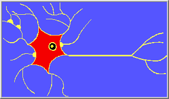
Neurons, commonly known as Nerve cells, largely constitute the Nervous System. They enable multicelled organisms to react to their environment and respond to its ever-changing conditions. Although their Axons can extend for many millimeters, Neurons can only be seen with proper staining techniques and a microscope that magnifes them to 40 times their size. But to visualize structures inside the Neuron, powerful electron microscopes are required that can magnify up to 10,000 times actual size. The Brain is an inticate network, consisting of approximately 200 billion Neurons and more than a trillion Glia Cells. Each Neuron receives and combines multiple inputs to determine, whether to transmit an action potential to the next target in its network (Neuron, Muscle, Gland, or Organ).
SubCellular Anatomy Functions Of NeuronsMetabolic machinery within the cell provides a power source, for information processing functions. In addition, the cell enforces a certain unity, for biochemical mechanisms throughout its extent. A tree of processes (extensions) called Dendrites is covered with special structures called Synapses, where junctions are formed with other Neurons. Synaptic contacts are the primary information processing elements in Neural Networks. Processes are similar to wires, conveying information over a finite distance. But, the resistance of finer Dendrites allows the electrical potential at their tips, to be computed with only partial coupling, to other computations on the Dendrite Tree. Temporal integration of signals occurs over the short term, through charge storage within the Capacitance of the cell membrane, and over the longer term, by means of internal second messengers and biochemical mechanisms. Neurons have long, specialized processes called Axons, which "digitalize" data for local transmission, and for transmitting data over long distances. The Axon is a hollow tube filled with fluid (AxoPlasma). Many Trophic substances made in the Neuron's Perikaryon are transported thru the Axon to the terminal buttons (End Feet), for further processing and packaging. Used NeuroTransmitters, Enzymes, and Trophic Factors are re-absorbed (Re-Uptake) by the End Feet, and transported back up the Axon to the Neuron for reusage. The flow process also provides cell components with necessary sugars, and other metabolites, needed for normal functions. The flow of substances in both directions is called AxoPlasmic Transport. The classic Neuron is equipped with a tree of filamentary Dendrites that algebraically add (the sum of positive and negative) elecrical Synaptic inputs from other Neurons. These input currents are integrated by the Capacitance of the cell membrane, until a Threshold Potential (all or nothing response) is reached. At which point an output is generated in the form of a Nerve Impulse - the Action Potential. This output Impulse propagates down the Axon, ending on another Neuron's dendrite tree of Synaptic contacts. The electrical resistance of a Nerve's CytoPlasm is sufficiently high that signals can not be propagated more than about 1 millimeter, before they would be spread out in time and their information lost. For this reason, Axons are equipped with an active amplification mechanism, which both periodically restores the Action Potential, and aids its continued propagation. In UnMyelinated Axons, this electric restoration is continuously done along the length of the Axon; however, this uses a lot of metabolic energy, and produces a fairly slow conduction speed. Many CNS Axons are segmentally wrapped by the extensions (Myelin), of many different Oligodendrocytes. This cellular cooperation, reduces the Capacitance between a Neuron's negatively charged Axonal Membrane (InterNode) and the surrounding positively charged ExtraCellular Fluid. Thus, Myelin greatly increases the conducting velocity of Action Potentials, while reducing the overall expenditure of Neuronal energy (ATP). The sheaths of Myelinated Axons have gaps called Nodes of Ranvier (Image) every few millimeters, where Sodium (Na+) and Potassium (K+) Ions are exchanged (Saltatory Conduction). Nodes of Ranvier are Ion channels that conduct electric current, where the Action Potential's Voltage is periodically restored. A single Myelinated Fiber can carry signals over a distance of 1 meter or more, at a speed of 150mm/msec. Whereas, UnMyelinated Nerve fibers conduct signals much more slowly - 20 to 30 mm per second. |
|
|
|
|
|
|
|
|
|