N
Necrosis - Is tissue decomposition resulting from the loss of its Blood and Oxygen supply, Burns, or other severe injuries. It can also be caused by some medications, commonly used to treat MS.
- Necrosis of the skin occurs after a subcutaneous injection, when the body is intolerant of the medication. Necrosis of the Hip and Shoulder Joints are caused by the Long-Term use of Steroids. #07
(See: Steroids)
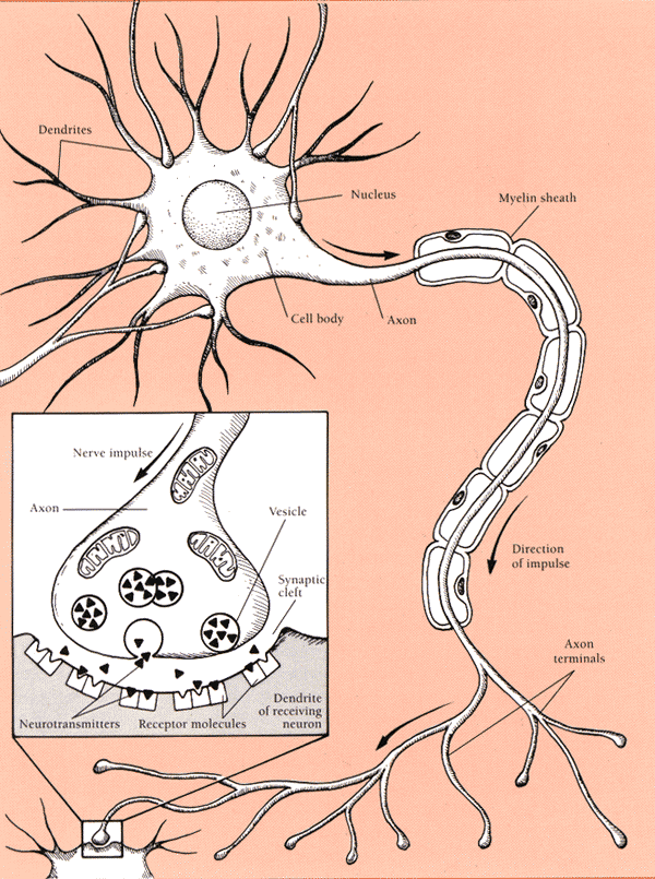
Nerve Fibers (Axons) - Are long, sparsely branched Processes, having non-changing diameters that extend from a Neuron's Cell Body and connect another Neuron's Axon, Dendrite, or Cell Body and/or bodily organs that compose their Neural Network. #25
- A bundle of Nerve Fibers (Axons) are either:
- Afferent - leading towards the higher Brain (CNS) and serving in the Perception of Sensory stimuli of the Skin, Joints, Muscles, and Inner Organs; or
- Efferent - leading away from the higher Brain and mediating contractions and relaxation of Muscles or Organs. #28
Neurologic
Disease - Any disorder of the Nervous System. There are many different Neurologic Diseases, among which is Multiple Sclerosis. #25
Neuron
- An individual Nerve Cell and the key data-processing cell of the Nervous System. Each has a Nucleus within a Cell Body and one or more Processes (Extensions) called Dendrites and Axons. #25, #28
NeuroTransmitters - Are chemicals (Small Molecules or Hormones), stored in small Synaptic Vessicles clustered at the tip of the Axon (terminal buttons). When a Nerve Impulse arrives for transmission to the next Neuron, they cross the Synapse enabling message transmission to another Neuron or the Stimulation of an Effector Cell (Muscle or Gland). (View: Image)
- When a NeuroTransmitter is received, it either Excites (Depolarizes) or Inhibits (Hyperpolarizes) the PostSynaptic Neuron. More than 30 organic molecules are thought to act as NeuroTransmitters.
- Some examples are: Acetylcholine, NorEpinephrine (precurser to Adrenaline), GABA, Serotonin, and Dopamine, although each acts in different responses. Some are Excitory, such as Acetylcholine, Serotonin (increased GastroIntestinal Motility), NorEpinephrine, and Dopamine; others are associated with Relaxation, such as Dopamine and Serotonin.
Neutrophils - A Phagocyte member of Leukocyte Cells, they are the Adaptive Immune System's first line of defense against Bacterial infections. After leaving nearby Blood Vessels, these cells follow chemicals produced by Bacteria in a cut or scratch and proceed to locate and eliminate the invader.
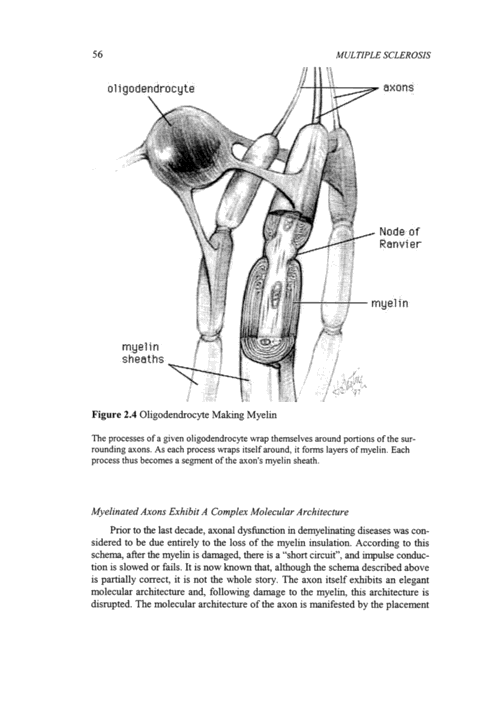
Nodes of Ranvier - Are the only gaps between Myelin sections (InterNodes) along Myelinated Axons, where Sodium (Na+) and Potassium (K+) can be exchanged (Saltatory Conduction); hence, continuing the Nerve signal's rapid transmission, to its target. They are crucial electrical refresher sites, where Action Potentials are restored. (View: Image) #20
Nuclear Magnetic Resonance (MRI, MR, NMR) - A diagnostic test that uses the magnetic properties of different substances in a Magnetic Field to produce images of the Brain, Spinal Cord, and other soft tissues of the Body. A MRI shows areas of Sclerosis (Lesion, Plaque), when they are larger than 2mm (Macroscopic Lesions).
- Scans can NOT show Microscopic Lesions, as they are too small for current imaging resolutions; but are included in your Lesion Load and Atrophy totals. These early smaller lesions are better documented, by Evoked Potential Tests, which are equally valid in meeting a Laboratory Supported Definite Multiple Sclerosis diagnosis.
- While this is the only test in which some Multiple Sclerosis Lesions can be seen, it cannot be regarded as conclusive; because, all lesions do not register on MRI scans and many other diseases can produce identical MRI images.
- MRI shows the size, quantity and distribution of Lesions larger than 2mm, and together with supporting evidence from your other diagnostic tests, Medical History, and Neurological Examination, may be a positive finding that confirms the MS diagnosis.
- It also provides an objective measure (Para-Clinical Evidence) of MS lesion activity in the Brain and Spinal Cord; however, Conventional MRI (T1 and T2 images) are NonSpecific (unknown cause) and have little relation to MS progression or Disability.
Magnetization Transfer and Proton MR Spectroscopy are two imaging techniques that better correlate with MS activity. They are not yet widely used, but newer more specific imaging protocols are presently being formulated. #25
- Abnormal MRI scans are found in
- 96% with a definite diagnosis of MS
- 70% with a diagnosis of probable MS
- 30 - 50% with a diagnosis of possible MS
- MRI Criteria for diagnosing MS
- At least 3 Lesions and two of the following:
- Lesions abutting the Lateral Ventricles
- Lesions with diameters greater than 5mm
- Lesions present in the Posterior Fossa (InfraTentorial)
Nocturia - Inability to hold urine while sleeping, resulting in bedwetting and/or disrupted sleep, due to repeated bathroom trips. (See: Neurogenic Bladder, Urgency with Hesitancy)
Nystagmus - A back and forth twitching Eye movement (Rhythmical jerking movements), with the fast component maximal, towards the side of the Cerebellar Lesion. Characterized by rapid, involuntary Eye movements, in the horizontal, or occasionally the vertical direction. #02, #25, #28
(Also See: Oscillopsia; InterNuclear Ophthalmoplegia; Optic Neuritis; Retrobulbar Neuritis)
|