My 7th MRI Report

3-19-2007 MRI at St. Peter's Hospital

MRI Brain WO/W Contrast


Clinical History: Multiple Sclerosis

Technical Factors:
Using a 1.5 Tesla superconducting magnet, T1 weighted and FLAIR sagittal and T1 weighted, FLAIR, and Diffusion weighted axial images were obtained.

Following intravenous administration of Gadolinium DTPA contrast, T1 weighted axial and coronal images were again obtained.

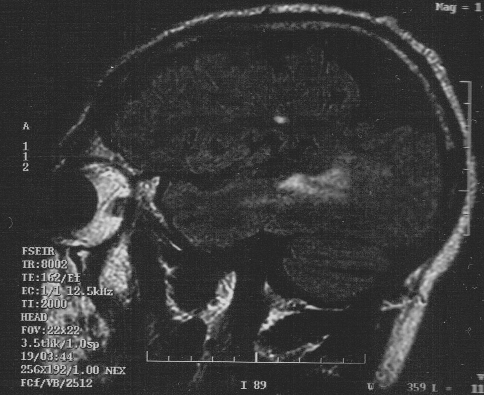
Comparison is made with previous MRI of the Brain of 11-11-2005

Findings:
The Ventricles are at the upper limits of normal in size. There is no evidence of Ventricular displacement.

There is localized Cortical Atrophy in the Parietal Lobes bilaterally, left more than right.

There are multiple areas of increased T2 signal in the PeriVentricular White Matter of both Cerebral Hemispheres.

There is a small area of increased T2 signal in the left Cerebellar Hemisphere as well.

These findings are unchanged from the previous MRI scan obtained at Albany Advanced Imaging on 11/11/05.

There is no evidence of abnormal contrast enhancement or restricted Diffusion.

There is no evidence of mass effect or ExtraCerebral fluid collection.

    Impression:

  1. No change compared to previous MRI of the Brain obtained at Albany Advanced Imaging on 11/11/05.
  2. Multiple White Matter T2 HyperIntensities compatible with the clinical diagnosis of Multiple Sclerosis.
  3. Localized bilateral posterior Parietal Lobe Cortical Atrophy is again noted, worse on the left. (View: Image)


Medical Texts
Anatomy | Immune System | Lymphocytes | Meds
MHC | Movement | Cranial Nerves | Physiology


MS Glossary ThJuland's MSers' Glen - Our CyberHome Page Top The Glen's Gallery: Come & Share Our Stories MS Files MS Abstracts Site Index


Abstracts
ANS | Bladder | Cognition | Fatigue | Fluid | Genetics
Interferons | IVIG | Nitric Oxide | Optic Neuritis | Pain
Physiology | Prions | Prognosis | ReMyelinate | Steroids
Stress | Treatments | TNF | Uric Acid | Viruses


© Copyright 1997 - 2011:
Permission is granted to MS Societies and all MSers to utilize information from these pages provided that no financial reward is gained and attribution is given to the author/s.