My 8th MRI Report

1-06-2009 MRI at St. Peter's Hospital

MRI Brain WO/W Contrast


Clinical History: Multiple Sclerosis

    Technical Factors:

  1. Using a 1.5 Tesla superconducting magnet, Multi-planar, multi-sequence MR imaging of the Brain was performed before and after the administration of contrast.

Comparison with Brain MRI dated 3/19/07.

    Findings:

  1. There is no intra or extra Axial hemorrhange.
  2. The Ventricles, Cisterns, and Sulci are prominent consistent with Atrophy.

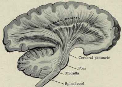
  3. Again are seen multiple focal areas of T2 and FLAIR HyperIntensities within the Corona Radiata (View: Image) and Centrum Semiovale of both Cerebral hemispheres.
  4. This would be consistent with patient's clinical history of Multiple Sclerosis.

  5. There are no abnormal areas of enhancement. There is no restricted Diffusion.

    Impression:

  1. Multiple White Matter T2 HyperIntensities compatible with clinical diagnosis of Multiple Sclerosis. Findings are unchanged when compared to prior examination.
  2. Localized bilateral posterior Parietal Lobes, Cortical Atrophy, left greater than right, unchanged.


Medical Texts
Anatomy | Immune System | Lymphocytes | Meds
MHC | Movement | Cranial Nerves | Physiology


MS Glossary ThJuland's MSers' Glen - Our CyberHome Page Top The Glen's Gallery: Come & Share Our Stories MS Files MS Abstracts Site Index


Abstracts
ANS | Bladder | Cognition | Fatigue | Fluid | Genetics
Interferons | IVIG | Nitric Oxide | Optic Neuritis | Pain
Physiology | Prions | Prognosis | ReMyelinate | Steroids
Stress | Treatments | TNF | Uric Acid | Viruses


© Copyright 1997 - 2011:
Permission is granted to MS Societies and all MSers to utilize information from these pages provided that no financial reward is gained and attribution is given to the author/s.