Findings:
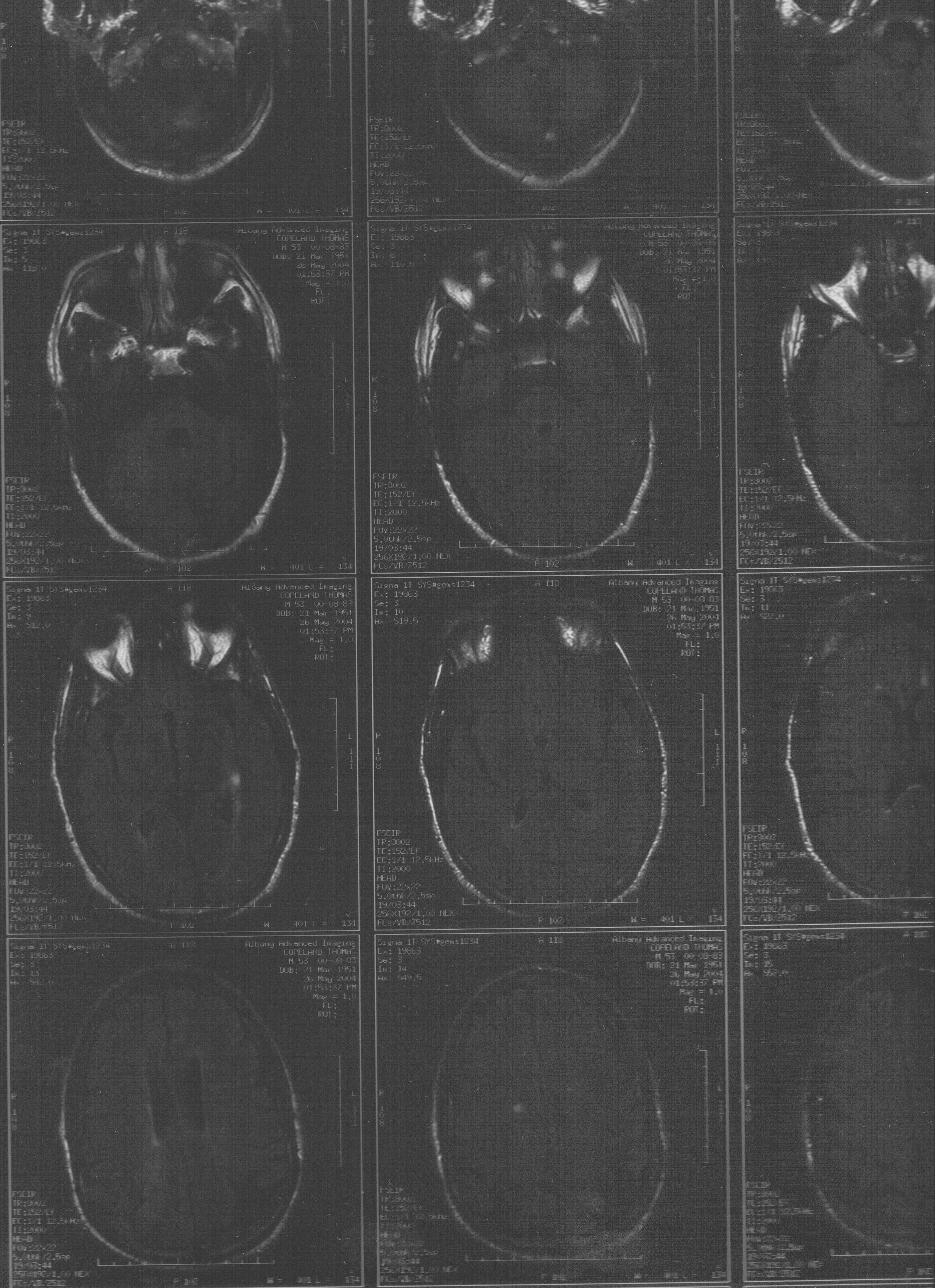
- There is no intra- or extra-axial hemorrhage. The Ventricles, Cisterns, and Sulci are prominent consistent with Atrophy, there is no evidence for mass or midline shift.
- There is no region of acute infarct or abnormal area of enhancement.
- Again are seen multiple focal areas of T2 and FLAIR HyperIntensity within the PeriVentricular White Matter of both Cerebral Hemispheres.
- Additionally, there is again demonstration of more diffuse hazy area of T2 and FLAIR HyperIntensity within the PeriVentricular White Matter.
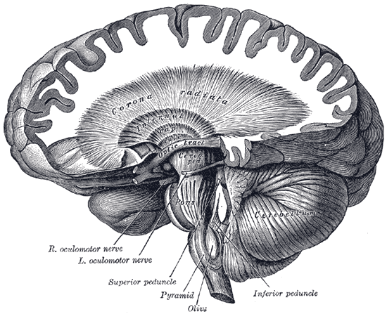
- Two dominant focal areas of signal abnormality are seen within the Right Corona Radiata. The more posterior area is not as apparent on today's examination. (View: Image)
- A few tiny focal areas are seen within the Left Corona Radiata which are mildly more prominent than prior study.
- Additionally, there is an area of signal abnormality involving the PeriVentricular White Matter, anterior to the Anterior Horn of the Left Lateral Ventricle which is more prominent than prior examination.
- None of these areas demonstrate abnormal enhancement after contrast administration.